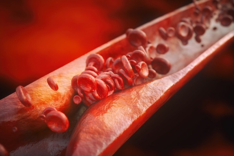
A chronic inflammatory condition of the arteries is atherosclerosis. Atherosclerotic plaque seen in the inner layer of arteries serves as a defining characteristic. Strokes or heart attacks can occur as a result of ruptured plaques. Substantial innate immune cells have been found to contribute to atherosclerosis.
Moreover, several T cell subtypes, including as CD8+ T cells and CD4+ regulatory T (Treg) cells, either promote or repress the sickness in mice. Yet, fundamental issues with T cell immunity identified in atherosclerosis remain unresolved. It is not known, in instance, whether T cell responses associated with atherosclerosis occur naturally in the bloodstream.
In the current study, researchers looked into tolerance checkpoints found in mice with advanced atherosclerosis, including atherosclerotic plaques, lymph nodes, and tertiary lymphoid organs.
The team employed aged Apoe -/- mice to examine atherosclerotic plaques, adventitial artery tertiary lymphoid organs (ATLOs), aorta-draining renal lymph nodes (RLNs), and the circulatory system in mice with atherosclerosis. RLN T cells from wild-type (WT) mice’s blood and were sequenced using paired 5′ single-cell ribonucleic acid-sequencing as controls (scRNA-seq). 13,800 T cells were used to generate a transcriptome repertoire atlas and T cell antigen receptor (TCR) atlas.Ten T-cell subgroups were identified in t-distributed stochastic neighbor embedding (t-SNE) plots based on their key T-cell markers and differentially expressed genes (DEGs). Plaques also comprised significant subsets of T lymphocytes and myeloid cells. Plaque T cell subgroups displayed pronounced aberrant properties in contrast to ATLOS, WT RLNs, and Apoe-/- RLNs. Plaques had more CD8+ Tem (T cell effector memory), CD8+ Tcm (T cell central memory), and -T cells and less Treg cells and naive CD4/CD8+ T cells.
Using flow cytometry, the scientists further confirmed that the proportion of CD4+ and CD8+ Tem cells in ATLOs increased in comparison to Apoe-/- secondary lymphoid organs (SLOs). Importantly, the percentage of naive CD4+/CD8+ T lymphocytes was lower in plaques.


